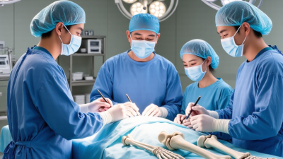
Bone is living tissue, not a fixed building material. It changes with age, pressure, disease, medication, and injury. That is why bone preservation techniques matter in modern orthopedic, dental, and reconstructive care. They aim to protect existing bone before damage becomes difficult to reverse.
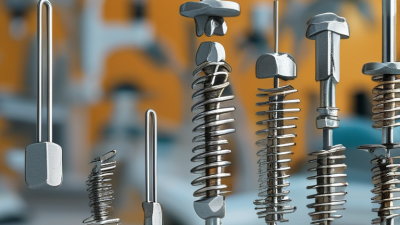
Harold M. Frost, MD, a leading researcher in skeletal biology, stated, “Bone modeling and remodeling are not the same thing.” This distinction remains important. Remodeling replaces older tissue, while modeling can change bone size and strength. Clinicians use this knowledge when planning atraumatic surgery, implant placement, fracture care, and grafting procedures. Small choices matter. A cooled surgical site, careful instrument handling, and stable fixation may help reduce unnecessary bone loss. A preserved ridge can also provide better support for a future dental implant.
The benefits are practical, but they are not guaranteed. Bone quality varies between patients. Smoking, diabetes, osteoporosis, infection, and poor nutrition can weaken outcomes. Even advanced methods cannot restore every millimeter of lost tissue. That limitation deserves honest attention. Bone preservation techniques should support biological healing, not replace sound diagnosis or experienced clinical judgment.
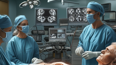
A strong treatment plan therefore begins with imaging, medical history, and realistic goals. It also requires monitoring after treatment. The visible result may look successful, while deeper bone changes continue quietly. Long-term follow-up can reveal those problems early. In this field, preservation is less about perfection and more about protecting future options. That perspective can improve function, reduce unnecessary procedures, and help patients retain healthy bone for longer.
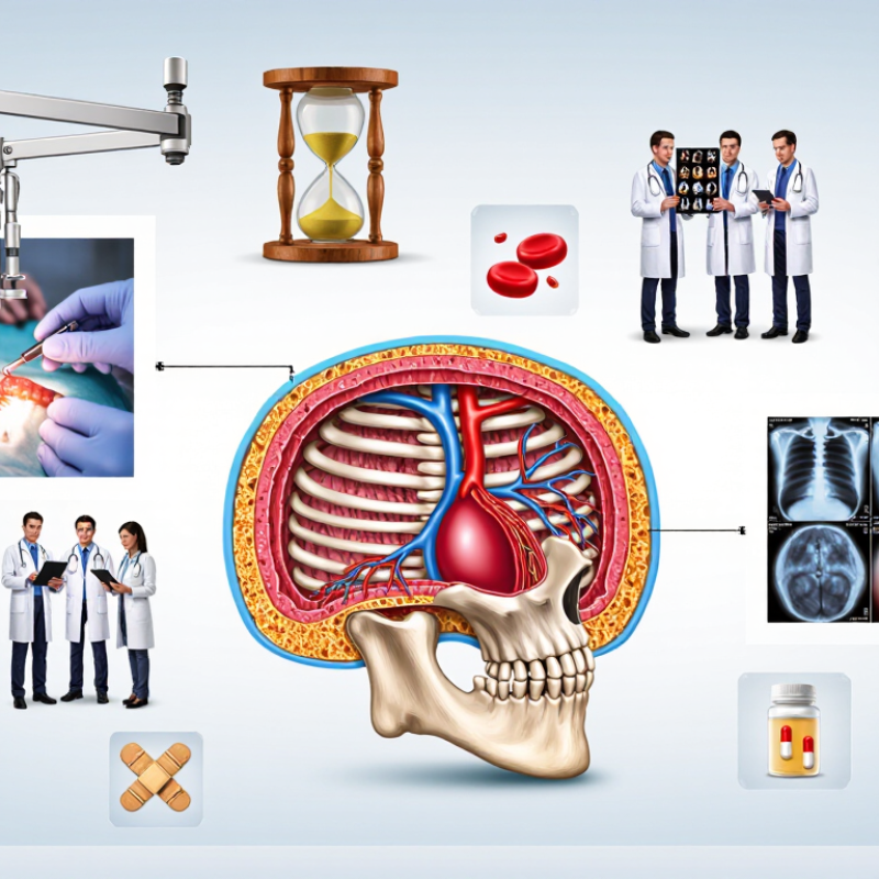
Bone preservation is a dental procedure designed to limit bone loss after a tooth is removed.
The jawbone needs stimulation from natural tooth roots. Without that pressure, the surrounding bone can gradually shrink in height and width. This change may affect chewing, facial support, and future implant planning.
The process usually begins with careful removal of the tooth and cleaning of the empty socket.
A clinician may place a biocompatible graft material inside the space. A protective membrane can cover the area and help stabilize the graft. The body then grows new tissue through the site over several months.
Healing time varies. Smoking, infection, diabetes, and poor oral hygiene may slow it down.
It is not a magic shield.
Bone preservation can reduce resorption, but it cannot prevent every change. The result depends on the socket’s condition, the patient’s health, and the technique used.
Clinical examination and dental imaging help determine whether preservation is suitable. Patients should also understand that graft material does not instantly become natural bone. That expectation can create disappointment.
Follow-up visits matter because clinicians need to check healing, membrane exposure, and signs of infection.
Mild swelling is common, but increasing pain, fever, or persistent bleeding needs prompt professional attention.
Bone preservation protects more than bone volume. It supports movement, balance, chewing, speech, and joint stability. When bone weakens or shrinks, nearby tissues may also lose support. A missing tooth, for example, can reduce stimulation in the jawbone over time. The change may be gradual, but the effects can become visible in facial contours and bite function.
Preservation may involve treating infection, repairing damaged bone, reducing unnecessary surgical trauma, or maintaining healthy loading through safe movement. In orthopedic care, clinicians may assess nutrition, fracture risk, muscle strength, and fall patterns. In dental care, they may evaluate bone levels with clinical examinations and appropriate imaging. These decisions should reflect age, medical history, healing capacity, and daily demands. One method does not suit everyone.
No technique guarantees perfect results. Biology is less predictable than a treatment plan suggests. Healing can vary with smoking exposure, diabetes control, medication use, and local blood supply. A common mistake is focusing only on the procedure while ignoring sleep, protein intake, or rehabilitation. That view is incomplete. Good care requires monitoring changes over time, explaining realistic risks, and adjusting treatment when healing does not follow expectations. Small choices matter. A stable walking routine, timely dental care, and professional evaluation after persistent pain can help protect structure and function.
Bone loss rarely begins with a dramatic warning. Aging slows bone remodeling, while menopause can accelerate mineral loss through declining estrogen. The International Osteoporosis Foundation reports that one in three women and one in five men over 50 will experience an osteoporotic fracture. That risk is not abstract. A weakened hip may turn a short fall into months of limited movement.
Lifestyle and medical factors also reshape bone structure. Long periods of inactivity reduce mechanical stimulation. Low calcium or vitamin D intake can impair mineralization. Smoking and heavy alcohol use may further reduce bone quality. Long-term corticosteroid therapy is another recognized cause.
In oral bone, untreated gum inflammation can destroy the tissue supporting teeth. Infection, trauma, and delayed treatment may leave uneven surfaces, cavities, or fragile edges.
Tips: Include resistance exercise and balance training when medically appropriate. Discuss calcium, vitamin D, and medication risks with a qualified clinician. After a fracture or tooth loss, early assessment matters. The National Osteoporosis Foundation estimates that about 10 million Americans have osteoporosis, while another 44 million have low bone density. Yet numbers cannot predict every patient. A scan may look acceptable, but pain, poor balance, or previous fractures still deserve attention. Preservation is not only about density; it is about keeping bone strong enough for daily demands.
Why Are Bone Preservation Techniques Important?
Medical techniques used to preserve bone help maintain tissue for reconstruction, research, and carefully selected treatments. The process begins with donor screening, informed consent, and strict infection testing. During collection, surgeons protect the bone from unnecessary damage. Small samples are handled in sterile conditions, with clear records from donor to storage.
Deep freezing is widely used for many structural grafts. Low temperatures slow cellular breakdown and preserve the bone’s shape. Freeze-drying removes moisture and can make storage and transport easier. Some grafts are also processed to reduce immune reactions. However, processing may affect strength, flexibility, or biological activity. The best method depends on the graft’s purpose.
A preserved bone graft may support a damaged joint, fill a cavity, or guide new bone growth. Surgeons examine its size, density, sterility, and storage history before use. Tissue banks monitor temperature continuously because one unnoticed failure can compromise an entire batch. Radiation may reduce contamination, but excessive exposure can weaken the graft. No method is flawless.
Clinical judgment remains essential. A technically preserved graft may still be unsuitable for a particular patient. Healing also depends on blood supply, stability, age, and overall health. Medical teams should explain these limits clearly, rather than presenting preservation as a guarantee. Better documentation and longer follow-up could improve future decisions.
Bone preservation helps maintain structural integrity, reduce microbial contamination, and retain biological properties for reconstructive surgery. The chart shows representative storage temperatures used for commonly preserved bone-tissue formats; exact protocols vary by tissue bank and regulatory requirements.
Lower temperatures slow enzymatic activity and cellular degradation. Fresh-frozen and cryopreserved tissues require highly controlled cold-chain storage, while freeze-dried bone can generally be stored at controlled room temperature when sealed and processed according to validated tissue-bank procedures.
Bone preservation matters because healing changes the shape and strength of the jaw. After a tooth is removed, surrounding bone may shrink in width and height. This loss can affect chewing, facial support, and future restorative options. Socket preservation, often using a carefully selected graft, helps maintain space while the body repairs the area. It does not create instant new bone. Healing takes time.
A stable foundation can support more predictable recovery. It may help a clinician plan a future implant or removable restoration with fewer adjustments. It can also reduce the chance of a sunken gum contour near the missing tooth. Recovery still depends on overall health, smoking status, infection control, and daily aftercare. Gentle cleaning, prescribed medication, and follow-up visits matter. Small details matter.
Evidence-based care begins with an examination and suitable imaging. Clinicians should explain graft materials, healing expectations, costs, and possible complications before treatment. Preservation is not necessary for every extraction, and it cannot guarantee an implant or perfect appearance. That limitation deserves honest attention. Some patients heal more slowly than expected or need another procedure. Long-term well-being depends on realistic planning, healthy habits, and regular professional review.
Copyright © 2021 United Orthopedic Corporation All Rights Reserved.
Necessary cookies are absolutely essential for the website to function properly. This category only includes cookies that ensures basic functionalities and security features of the website. These cookies do not store any personal information.
Any cookies that may not be particularly necessary for the website to function and is used specifically to collect user personal data via analytics, ads, other embedded contents are termed as non-necessary cookies. It is mandatory to procure user consent prior to running these cookies on your website.